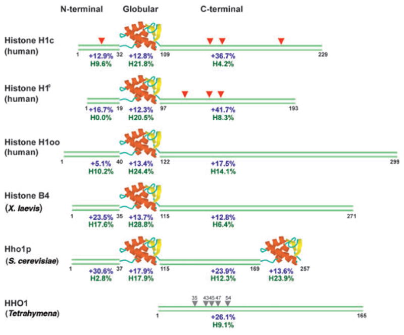
Figure 2.

Structural features of H1 variants. Human H1c represents the typical somatic variant found in most cells. The amino acid positions at the boundaries of the domains are indicated under each sequence. For each domain, percentage of net positively charged residues is indicated in blue and percentage of hydrophobic residues (Val, Leu, Ile) in green. Red triangles, conserved SPTXK motifs; gray triangles, serine residues in Tetrahymena HHO1 protein, which have been shown to affect binding49.
