
Figure 2.
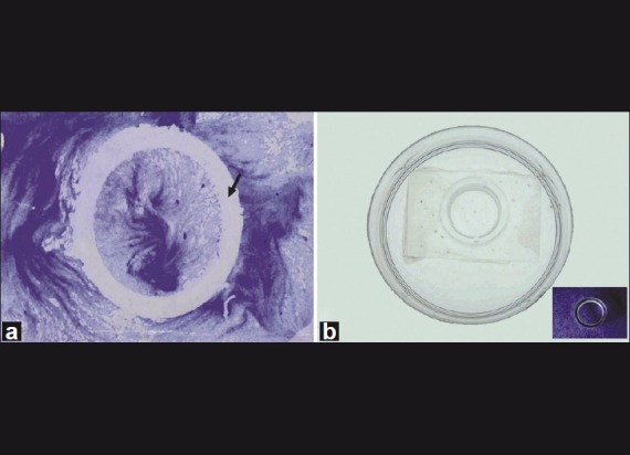
(a) Whole mount preparation of cultured epithelial cells on human amniotic membrane (barrier removed after culturing) showing the growth of limbal cells in the centre and conjunctival cells at the periphery outside the ring barrier and a clear circular zone, devoid of cells (arrow). [H and E, ×100], (b) Setup for the coculture of conjunctival and limbal tissues on a single HAM, on a Petri plate with limbal explants inside the ring barrier and conjunctival explants outside the barrier. (Inset: self-designed ring-shaped barrier made of Perspex)