
Figure 3.
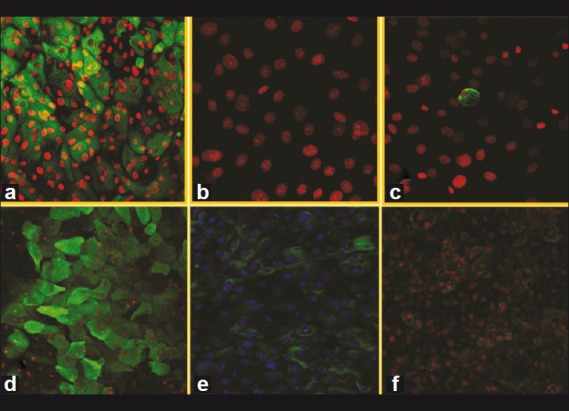
Conjunctival epithelial cells on human amniotic membrane showing (a) CK19 positivity with green (FITC labeled) cytoplasmic staining and red (PI labeled) nuclei [x200]. (b) CK3 negative with only red nuclei [×400]. (c) Presence of goblet cells positive for MUC5AC as shown by green cytoplasmic staining and red nuclei [×200] Limbal epithelial cells showing. (d) CK3 positivity with green cytoplasmic staining and red nuclei [×200]. (e) CK14 positive with green cytoplasmic staining and blue (PI labeled) nuclei [×200]. (f) Positive ABCG2 with green membrane staining and red nuclei; FITC = Fluorescein iso thio cyanide, PI = Propidium iodide