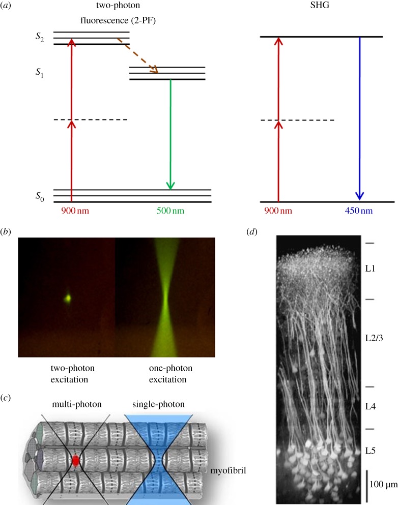
Figure 3.
Multiphoton microscopy: principle, micrometre-scale excitation and tissue penetration. In (a), the principle of 2-PF and SHG is explained, and the Jablonski diagrams show the energy levels involved and the excitation and emission processes. With 2-PF, the excitation does not occur with one photon of 450 nm, but with two near-simultaneous photons of 900 nm (half the energy) each. Owing to certain quantum mechanical selection rules that apply for 2-PF other than 1-PF, in reality, excitation energies are not exactly one-half (emission is unchanged). In SHG, two photons (900 nm) simultaneously striking a highly ordered, non-centrosymmetric material may induce a virtual energy state transition that lasts only as long as the illuminating pulse (approx. 1 fs) and results in rapid relaxation to S0 and the release of a photon of exactly twice the energy of the incoming photons (450 nm). No photons are absorbed, and resonance enhancement of SHG can occur. (b) Comparison of two- and one-photon excitation by use of a fluorophore solution. Two-photon excitation generates fluorescence exclusively in the focal volume, whereas in one-photon excitation, fluorescence is generated all along the light path. (c) Only with multiphoton fluorescence microscopy, is it possible to excite a confined focal volume (red) within a tissue or even cell. (d) Illustration of deep-tissue imaging in the mouse neocortex. A maximum intensity side projection of a fluorescence image stack obtained in a transgenic mouse expressing a genetically encoded indicator is shown. Nearly the entire depth of the neocortex can be imaged [27].

