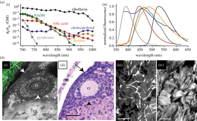
Figure 4.
Sources of intra- and extracellular autofluorescence. (a) Most important intracellular autofluorescent molecules are listed together with their two-photon absorption probability (two-photon action cross-section, (i) and emission spectra (ii)). AF can be excited over a large range of the visible spectrum. (b) AF is a suitable tool to visualize important tissue structures. (i) and (ii) show intracellular fluorescence sources: (i) presents an multiphoton image (fluorescence in greyscale, SHG in green-collagen) and (ii) the corresponding H&E-stained histological image of a mouse ovarium. The ovarian epithelium (arrow), oocyte (O), granulosa cells (arrowhead), thecal cells (T), the corpus luteum (CL) and ovarian bursa (OB) are all clearly resolvable and resemble the histological image in (ii). In (iii) and (iv), ECM structures are shown. Image of elastin fibres of a human skin explant ((iii), 740 nm) and SHG image (iv) shows collagen structure taken at 800 nm. Images from Zipfel et al. [108].

