Figure 6.
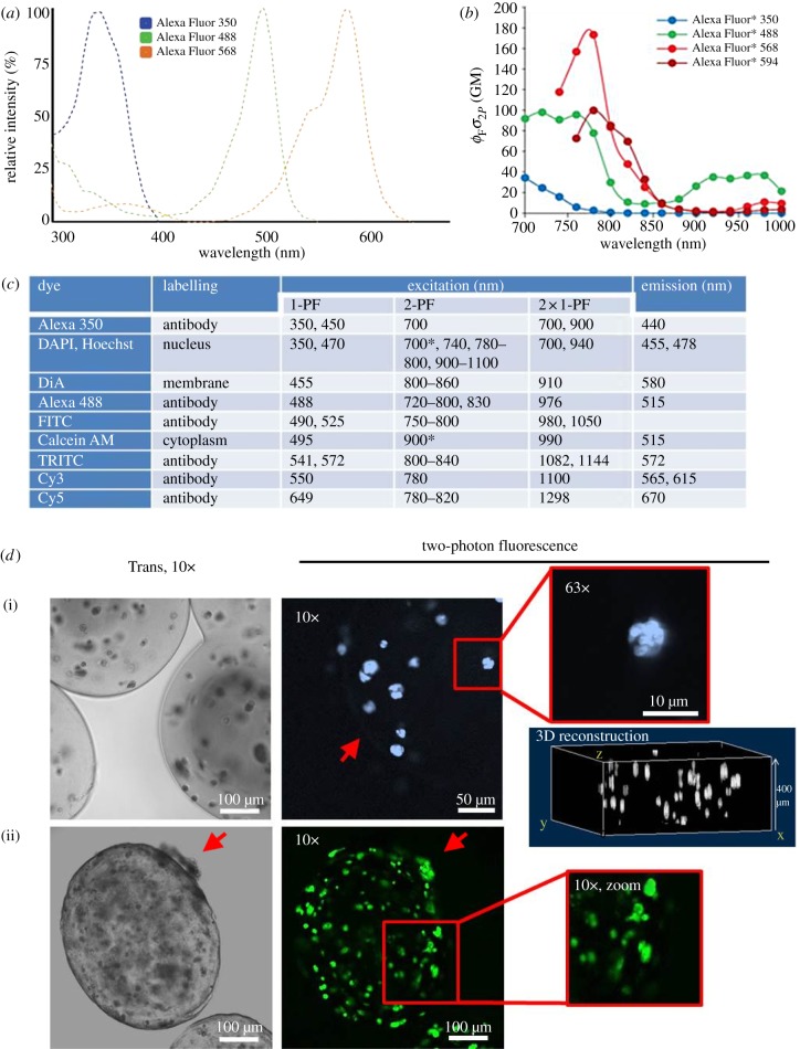
Two-photon excitation and imaging of selected fluorescent dyes. (a) Normalized one-photon excitation spectra of antibody-conjugated Alexa Fluor 350, 488 and 568 dyes are shown (unconjugated dyes almost identical). Spectra are narrow and well separated. Data from Spectra Viewer software (Invitrogen). (b) Two-photon excitation spectra of Alexa Fluor 350, 488, 568 and 594 dyes. The y-axis unit Goeppert–Mayer (product of fluorescence quantum yield ΦF and two-photon absorption cross section σ) is a measure of excitation efficiency. These spectra are shaped differently (less well defined and much broader) compared with (A, e.g. Alexa Fluor 488) and maxima are blue-shifted [100,108]. (c) Excitation and emission of prominent dyes using one- and two-photon excitation. The table lists a number of commonly used fluorophores (sorted by increasing excitation wavelength). Two-photon excitation maxima are shifted markedly to shorter wavelengths compared with double the wavelengths of one-photon excitation. Emission spectra, however, are identical. Sources: Zeiss publication ‘suitable dyes for multiphoton’, Xu et al. [121], 1-PF values from Spectra Viewer (Invitrogen). Asterisk denotes own observations. Alternatives to Cy and Alexa Fluor dye series with optimized photophysical behaviour are Atto, Chromeo and DyLight dyes. (d) Fluorescence-labelled encapsulated MG-63 cells imaged with 2-PF. (i) Cells in alginate-RGD capsules cultured for four weeks were DAPI-stained for nuclei and imaged at 700 nm. The red arrow points to the capsule border (10× image). Nuclear staining even resolved subnuclear segmentation (63×). A three-dimensional reconstruction image generated from image stacks allows quantitative analyses of cell number and distribution. (ii) Calcein-stained cells in alginate cross-linked with gelatin (50 : 50) after six weeks (900 nm). Capsules show a rough and porous surface and are partially non-transparent. Cells migrate through the border to the outer surface of the capsule (red arrows). The cellular calcein distribution allows determination of cell morphology (zoom image). 2-PF allows imaging deep within widely non-transparent capsules.

