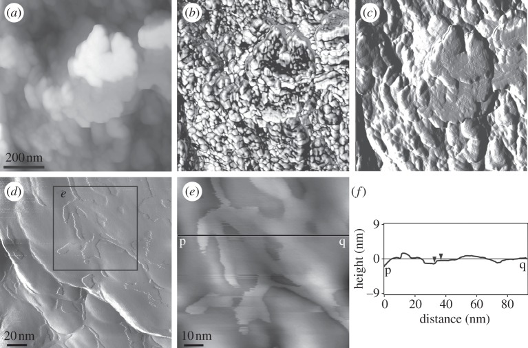
Figure 7.
AFM images of a vertical polished section through the outer prismatic layer of Pinctada margaritifera. The images were taken in tapping mode. (a–c) Height, phase and amplitude images, showing the nanoblocky structure. The scale bar in (a) is valid for the three images. (d) Close-up view (amplitude image) of the nanounits showing the membrane that covers the nanounits. (e) Detail of (d) (position indicated). Height image. (f) Height profile along the line p–q in (e). The vertical distance between the two triangular markers (i.e. the approximate thickness of the membrane shown in (e)) is 0.8 nm.

