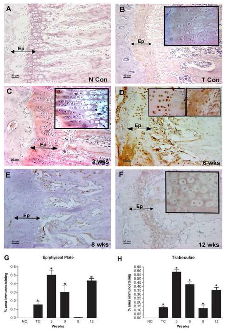
Figure 1.

A–F: Immunolocalization of PLF in the forelimb epiphyseal plate of radii and ulna of normal control (N Con), Trained control (T Con), or HRHF task (3, 6, 8, and 12 wks) animals. Forelimb bone sections were immunoreacted with PLF antibody (brown color indicates positive staining) and counterstained with haematoxylin (a nuclear stain). PLF was (A) not detected in bones of N Con animals as indicated by the lack of brown color, but (B) was detected at low levels in T Con animals in trabeculae and in proliferating, prehypertrophy, and hypertrophy zones of the epiphyseal plate (inset: cells in the epiphyseal plate shown at higher magnification, staining detected in the matrix surrounding cells). (C) PLF was detected at 3 wks in trabeculae and proliferating, prehypertrophy, and hypertrophy zones in the epiphyseal plate, and (D) at 6 wks in the nuclei of prehypertrophy chondrocytes (left and right inset), and in the matrix around hypertrophied chondrocytes (right inset) in the epiphyseal plate. (E) PLF was not detected at 8 wks in the epiphyseal plate, but (F) was detected at 12 wks in hypertrophied chondrocytes in the epiphyseal plate (inset). G–H: PLF levels in the epiphyseal plate (G) and trabeculae (H) was quantified using a microscope interfaced with an image analysis system (Bioquant Osteo II) and plotted as percent area of PLF immunostaining against weeks of task performance. Ep=epiphyseal plate, NCon/NC = normal control, Tb=trabeculae, TC = trained control.
