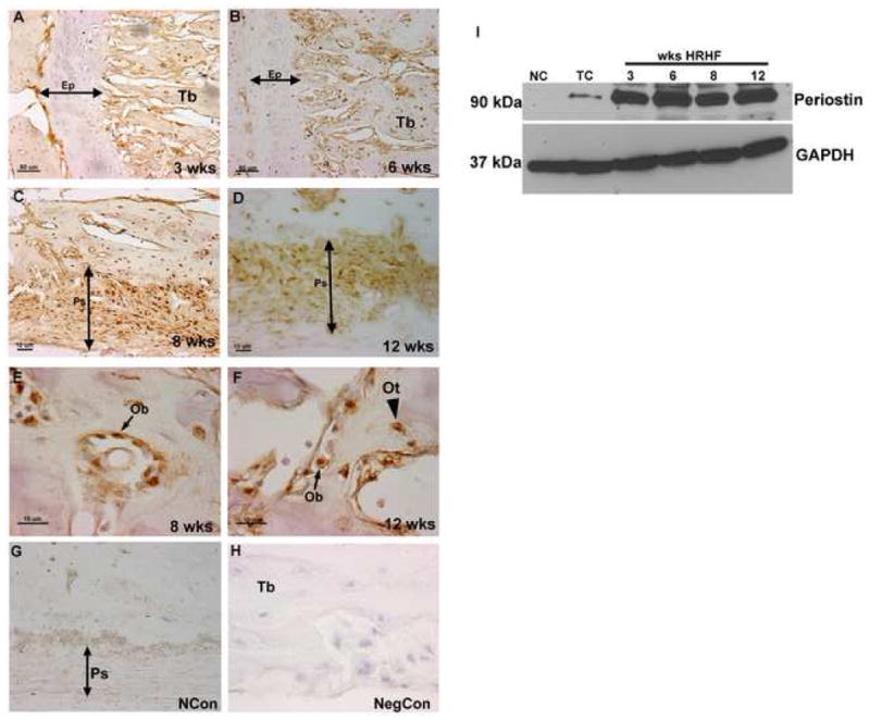
Figure 5.

A–I Immunolocalization of Periostin in the epiphyseal plate and periosteum and in osteoblasts and osteocytes of HRHF rats (3, 6, 8, and 12 wks). Forelimb bone sections (radii and ulna) were immunoreacted with Periostin antibody (brown color) and counterstained with haematoxylin. Periostin was detected in the periosteum, osteoblasts and osteocytes at all time points, but was not detected in the epiphyseal plate. Representative data for epiphyseal plate at 3 and 6 wks (A, B), periosteum at 8 and 12 wks (C, D), and osteoblasts and osteocytes at 8 and 12 wks (E, F). Normal control (NCon) section reacted with anti-Periostin (G) and negative control (NegCon; no primary antibody, H). Detection of Periostin by western blot analysis (I). Ep=epiphyseal plate, Ps=periosteum, Tb=trabeculae, Ob=osteoblast, Ot=osteocyte.
