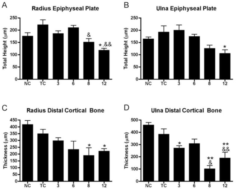
Figure 7.

A–D: Morphological quantification of epiphyseal plate height and cortical wall thickness of the distal radius and ulna of normal control (NC), trained control (TC), and HRHF (3, 6, 8 and 12 wk) animals. (A) Epiphyseal plate height was decreased in the distal radius in week 12 compared to normal controls (*: p<0.05), and in weeks 8 and 12 compared to trained controls (&: p<0.05; &&:p<0.01). (B) Epiphyseal plate height in the distal ulna was decreased in week 12 compared to normal controls (*: p<0.05). (C) Distal radius cortical thickness was decreased in weeks 8 and 12 compared to normal controls (*: p<0.05; **: p<0.01). (D) Distal ulna cortical thickness was decreased in weeks 3, 8 and 12 compared to normal controls (*: p<0.05; **: p<0.01), and compared to trained controls (&: p<0.05; &&: p<0.01).
