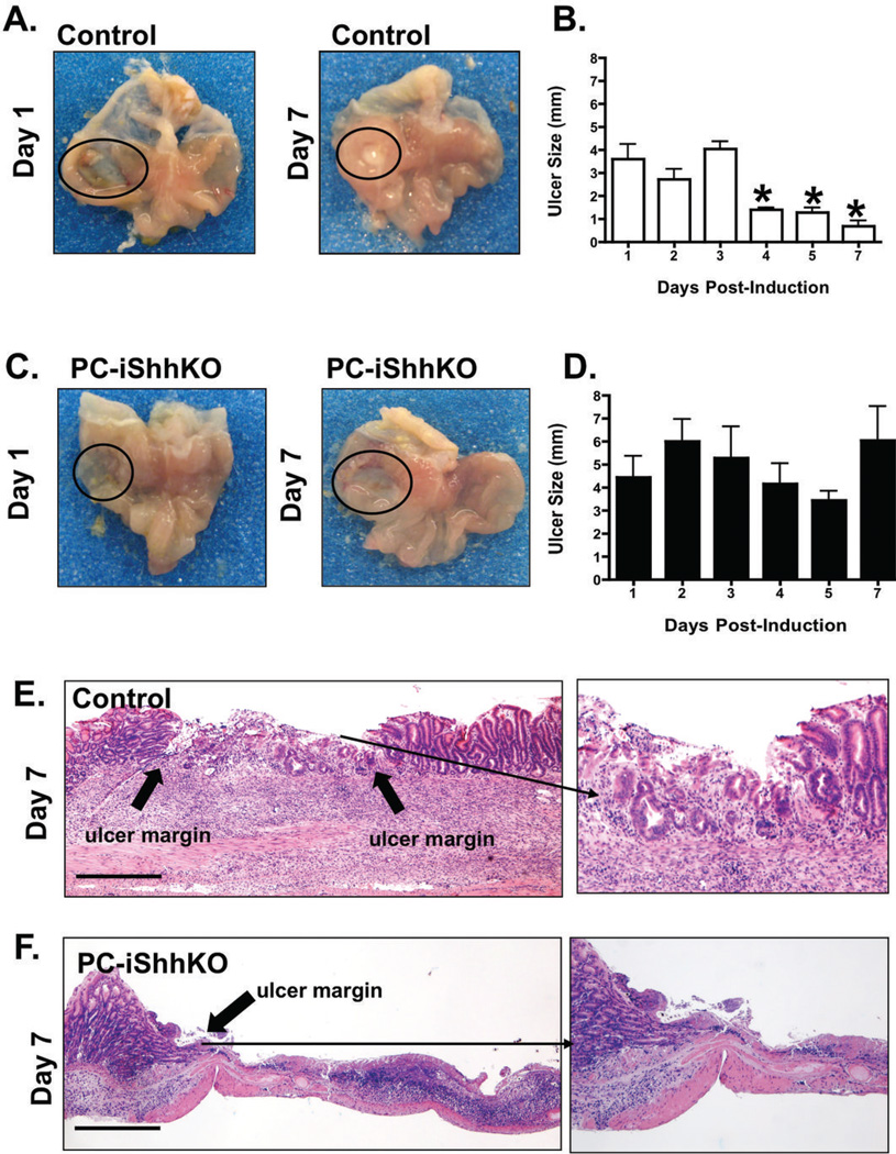
Figure 5. Wound healing in control and PC-iShhKO mouse stomachs.
Gross morphology and ulcer sizes measured in mouse stomachs collected from (A, B) control and (C, D) PC-iShhKO mice 1 to 7 days after acetic acid-ulcer induction. Ulcers are circled on the figures A and C. Data is shown as mean + SEM of ulcer size (mm). *P<0.05 compared to control group, n = 6–8 mice/group. H&E staining of stomach sections collected from (E) control and (F) PC-iShhKO mice 7 days after acetic acid ulcer injury. Block arrows show the ulcer margin and inset is a higher magnification of the shown area. Images were captured at 4X magnification, images shown in inset were captured at 10X magnification. Scale bars = 375 microns.

