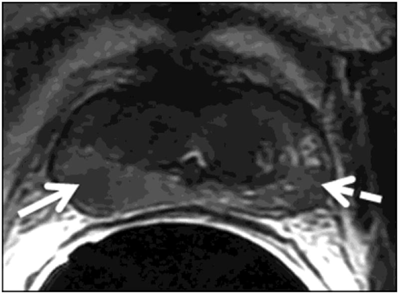
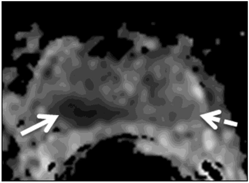
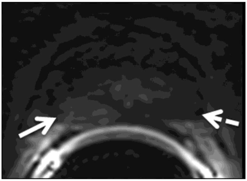
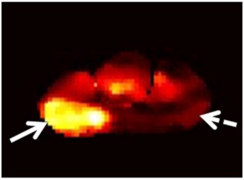
Figure 1.


A 75-year-old man with recurrent Gleason 4+3 tumor (arrows) after radiotherapy. An ill-defined lesion is noted in the right peripheral zone of the prostate on the T2-weighted image (1a), the tumor becomes more conspicuous on the ADC map (1b), dynamic contrast-enhanced image (1c) and Ktrans map (1d). Note that there is an ill-defined nodular area (dashed arrow) that could mimic tumor on the contralateral aspect of the prostate on the T2-weighted image (1a); however, ADC map (1b), dynamic contrast-enhanced image (1c) and Ktrans map (1d) rule out tumor on the left side (dashed arrows).
