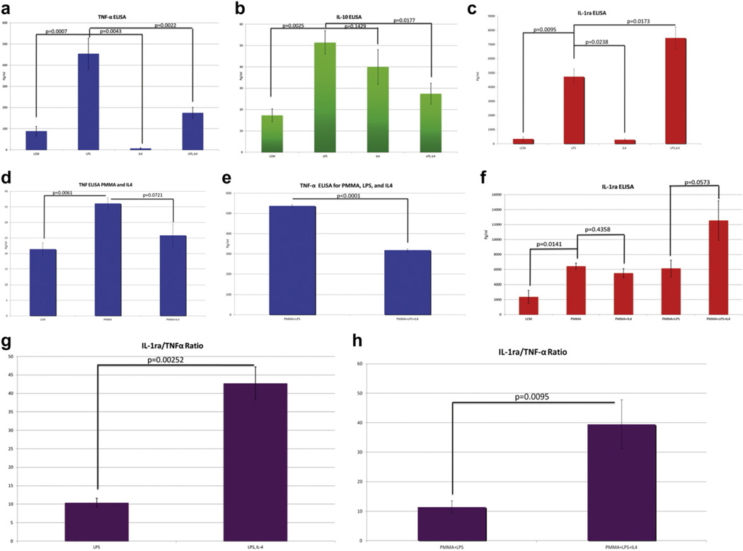
Fig. 4.
ELISA analysis was performed on cell culture supernatants taken from in vitro macrophage cultures stimulated with LPS with/without PMMA and IL-4. Analysis was performed for TNF-α, IL-10, and IL-1ra. A ratio of IL-1ra/TNF-α is also presented. (a) ELISA analysis of TNF-α in macrophages stimulated with LPS shows a higher level of section compared to conditioned media alone. This response is decreased with the addition of IL-4 after initial LPS administration. (b) ELISA analysis of IL-10 in macrophages stimulated with LPS shows a high level of secretion. This may be due to LPS induction of IL-10 production, which has been found to be a well-established effect. (c) ELISA analysis of IL-1ra in macrophages stimulated with LPS shows a high level of secretion. This response is increased by the subsequent addition of IL-4, suggesting that IL-4 administration following LPS priming may preferentially polarize macrophages towards M2, increasing the expression of IL-1ra. (d) ELISA analysis of TNF-α in macrophages stimulated with PMMA particles shows a significantly high level of section. This response is decreased with the addition of IL-4 after initial PMMA administration. (e) ELISA analysis of TNF-α in PMMA stimulated macrophages shows a significantly higher level of TNF-α release in response to PMMA particles with LPS administration. This response is decreased with the addition of IL-4 after initial PMMA and LPS priming. (f) ELISA analysis of IL-1ra macrophages stimulated with PMMA and PMMA with LPS priming prior to IL-4 administration shows a significantly high level of IL-1ra release. (g) The ratio of IL-1ra/TNF-α in the LPS only group was 1.20, whereas with LPS followed by IL-4, the IL-1ra/TNF-α increased to 4.46. This shows that the increase in IL-1ra expression is not based on TNF-α stimulation alone. (h) The ratio of IL-1ra/TNF-α in the PMMA with LPS group was 11.44, whereas with PMMA and LPS followed by IL-4, the IL-1ra/TNF-α increased to 39.47, showing that IL-1ra expression was increased beyond what would be expected if it was solely related to TNF-α release.

