
Fig. 6.
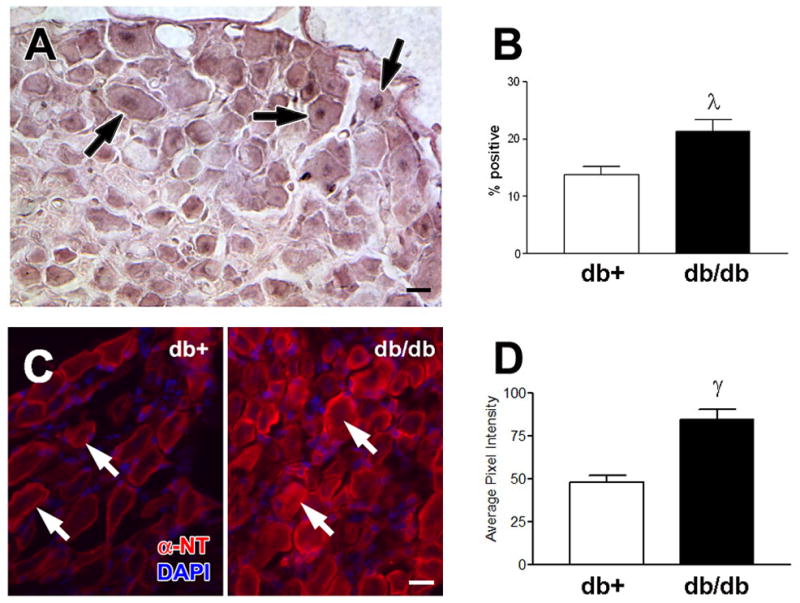
Damaged DNA was measured by TUNEL staining. A) TUNEL positive sensory neurons (arrows) were detected in the lumbar DRG of BKS-db/db. B) Increased number of TUNEL labeled DRG in BKS-db/db mice at 24 weeks, λp < 0.05. Five animals per group and > 150 neurons per animal were counted. Results are expressed as the percent TUNEL positive cells of total neurons counted. Localization of nitrated proteins was measured by nitrotyrosine immunofluorescence (NT- immunofluorescence). C) NT-immunofluorescence reveals an increase in nitrated proteins within DRG neurons (arrows) from BKS-db/db compared to BKS-db+ mice [nuclei stained with DAPI]. D) Histograms of the fluorescence signal indicate a relative increase in the intensity of NT-immunofluorescence in DRG from BKS-db+ versus BKS-db/db mice (γp<0.01). Bar = 20 μm. Open bars represent the nondiabetic measurements and the black bars represent the diabetic measurements.