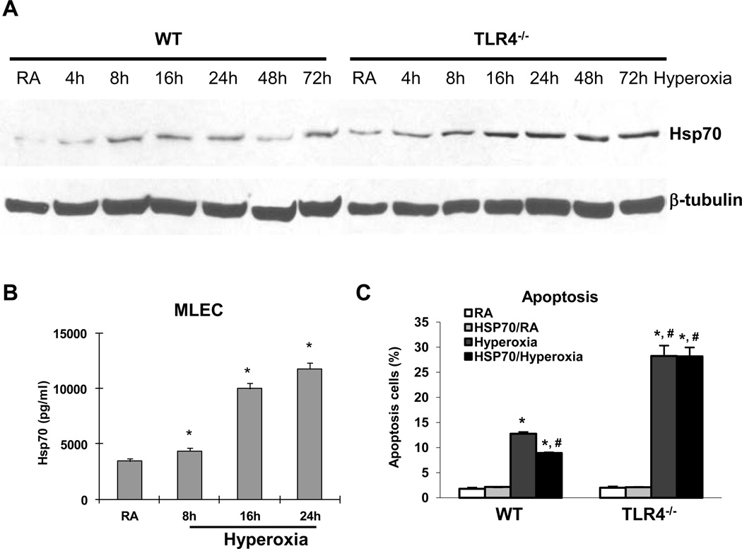
Figure 1. Hsp70 is inducted and secreted by MLEC during hyperoxia.
A) Hsp70 protein in WT and TLR4−/− MLEC by western blot after a time course of hyperoxia. β-tubulin as loading control. B) Hsp70 protein in WT MLEC supernatant was measured by ELISA after 8h, 16h, and 24h of hyperoxia. RA, room air control. * p <0.05 vs RA. C) WT and TLR4−/− MLEC were treated with recombinant human Hsp70 protein at 8.65µg/ml and were exposed to 72h of hyperoxia. RA, room air control. Graphical quantitation of flow cytometry analysis of apoptosis was determined. The values are expressed as mean ± SD. *p<0.05, vs RA WT; #p<0.05 vs hyperoxia WT (experiments were performed in triplicates).

