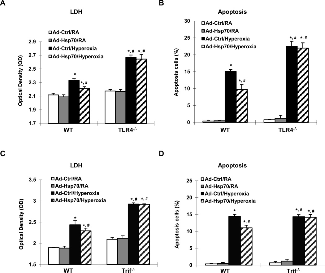
Figure 4. Hsp70 is protective and depends upon a TLR4-Trif pathway in MLEC.
WT, TLR4−/− and Trif−/− MLEC were treated with Ad-Ctrl or Ad-Hsp70 and were exposed to 72h of hyperoxia. RA, room air control. LDH activity of WT and TLR4−/− MLEC in (A), and WT and Trif−/− MLEC in (C) was determined. Graphical quantitation of flow cytometry analysis of apoptosis in WT and TLR4−/− MLEC in (B), and WT and Trif−/− MLEC in (D) was determined. The values are expressed as mean ± SD. *p<0.05, vs Ad-Ctrl/RA WT; #p<0.05 vs Ad-Ctrl/hyperoxia WT (experiments were performed in triplicates).

