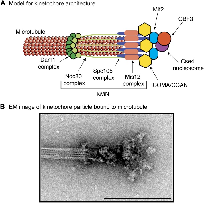
Figure 6.
Model for the budding yeast kinetochore. (A) Schematic indicating the rough position and stoichiometry of the budding yeast kinetochore subcomplexes. (B) Electron microscope image of a purified yeast kinetochore particle bound to a microtubule, originally published in Gonen et al. (2012). There is a ring that encircles the microtubule and globular domains that could represent KMN that touch the microtubule. Bar, 200 μm.

