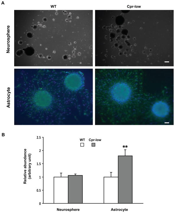
Fig. 2. In vitro formation of neurospheres and astrocytes from SVZ cells of Cpr-low and WT mice.
SVZ cells dissected from Cpr-low and WT mice (female, 6-month old, three per group) were examined in culture for their capacity to form neurospheres and astrocytes. A. Representative images of neurospheres (upper panel; dark field) originated from SVZ cells and astrocytes (lower panel; IHC) differentiated from neurospheres are shown. The astrocytes were detected using anti-GFAP; cells were also stained with DAPI. Scale bar: 200 μm for neurosphere, 50 μm for astrocyte. B. Numbers of neurospheres and astrocytes were counted and their relative abundance was calculated by setting the averaged numbers from WT group (94 and 36, respectively) as 1. Values represent means ± S.D., n=3. **, P<0.01; Cpr-low vs WT mice (Students t-test).

