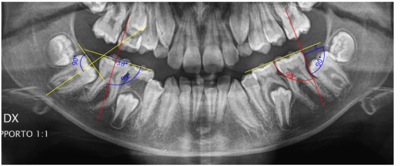
Figure 1.
Panoramic radiograph , with a magnification rate of 1:1 , at the time of one third of MM2 root formation (T1): The measurements of the angle of inclination of MM2 ( right side ) and of the distance from the distal height of the contour of the first mandibular molar (MM1) to the anterior margin of mandibular ramus (left side) are shown.

