
Figure 3.
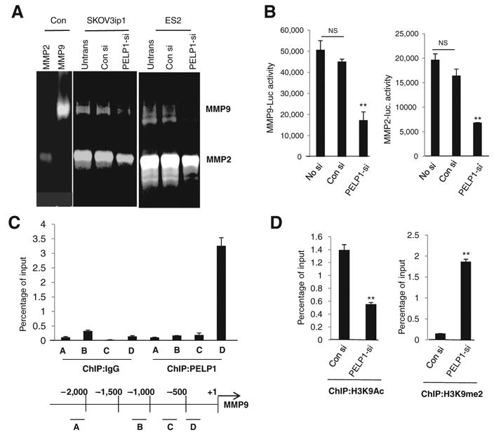
PELP1 regulates the expression and activities of MMPs. A, gelatin zymography analysis of activity of MMP2 and MMP9 in SKOV3ip1 and ES2 cells that were transfected with control siRNA (con si) or PELP1-siRNA (PELP1-si). B, SKOV3ip1 and ES2 cells transfected with MMP9-Luc and MMP2-Luc vectors, treated with control siRNA or PELP1-siRNA, and the reporter gene activity was measured after 72 hours. C, ChIP assay was done using the DNA isolated from SKOV3ip1 cells and by using antibodies specific for PELP1 or isotype rabbit IgG control. DNA recovered from ChIP or input controls was subjected to real-time qPCR using 4 primers (A, B, C, and D) that span the MMP9 promoter region. D, SKOV3ip1 cells were transfected with control or PELP1-siRNA and ChIP assay was done using antibodies specific for H3K9Ac or H3K9me2. DNA recovered from ChIP or input controls was subjected to real-time qPCR using primers that detect proximal MMP9 promoter region. The promoter occupancy was calculated on the basis of the ratio of ChIP/input control. **, P ≤ 0.001, t test. NS, nonsignificant.