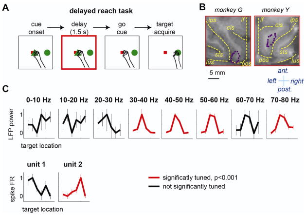
Figure 1.
A. Task sequence of the delayed reach task. The square and the triangle in the center represent the eye- and hand-fixation targets, respectively, and the circle in the periphery represents the reach target. B. The locations of 16 electrodes (purple dots) overlaid on the magnetic resonance image of each monkey’s brain. The yellow dashed lines indicate the intra-hemispheric fissure (if) and major sulci including the intra-parietal sulcus (ips), parieto-occipital sulcus (pos), lunate sulcus (lus), superior temporal sulcus (sts), and cingulate sulcus(cis). The anterior, posterior, left, and right directions are indicated. C. Tuning curves, the mean delay response for each of six target locations, of LFP and spike units recorded from the same electrode. The LFP power and spike firing rate (FR) in each tuning curve were normalized so that that the maximum is 1 and the minimum is 0. The error bars are the standard deviations.

