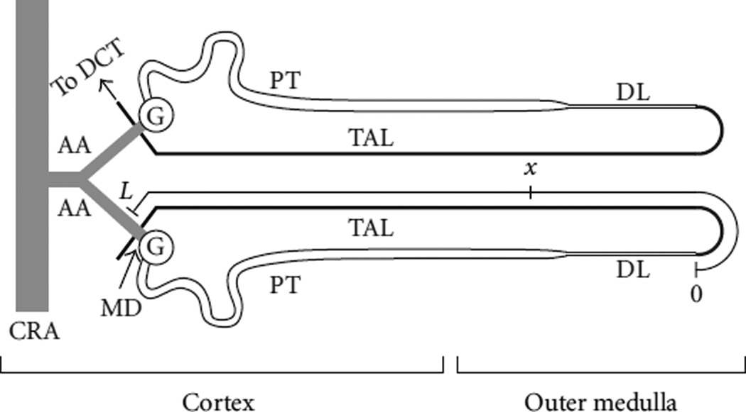
Figure 5.
A schematic drawing of two short-looped nephrons and their afferent arterioles (AA). The arterioles branch from a small connecting artery (unlabeled), which arises from a cortical radial artery (CRA). The nephron consists of the glomerulus (G) and a tubule having several segments, including the proximal tubule (PT), the descending limb (DL), the thick ascending limb (TAL), and the distal convoluted tubule (DCT). Each nephron has its glomerulus in the renal cortex, and each short-looped rat nephron has a loop that extends into the outer medulla of the kidney. The axis on the TAL of the lower nephron corresponds to the spatial axis used in the model; in this figure distance is indicated in terms of fractional (nondimensional) TAL length. Tubular fluid from the DL flows into the TAL lumen at x = 0; the chloride concentration of TAL luminal fluid is sensed by the macula densa (MD) at x = 1. The MD, a localized plaque of specialized cells, forms a portion of the TAL wall that is separated from the AA by a few layers of extraglomerular mesangial cells; in this figure, the MD is part of the short TAL segment that passes behind the AA. Fluid from the DCT enters the collecting duct system (not shown), from which urine ultimately emerges. Structures labeled on one nephron apply to both nephrons. (Figure and legend adapted from [14].)

