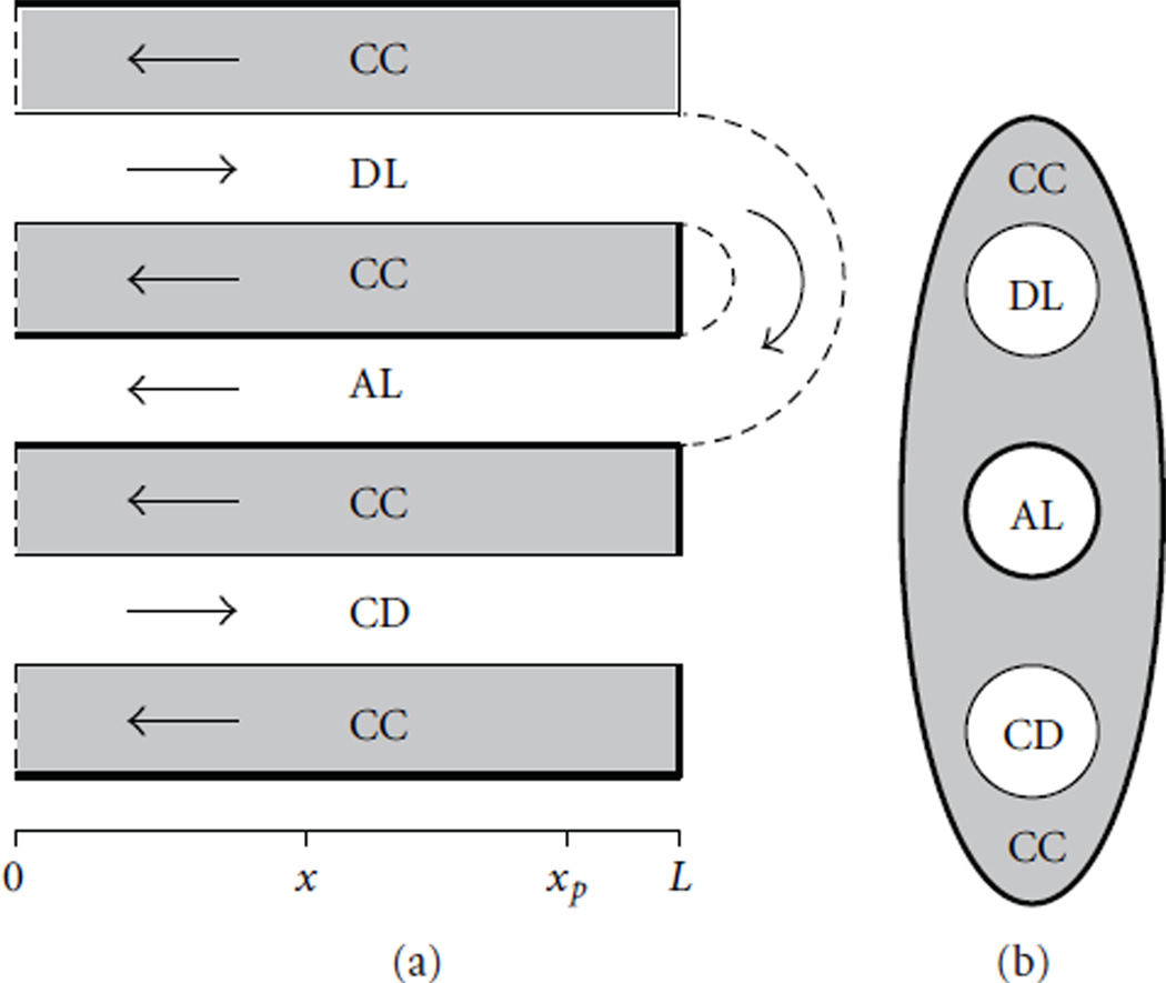
Figure 9.
Schematic diagram of the central core model. (a): Tubules along spatial axis. DL, descending limb; AL, ascending limb; CD, collecting duct; CC, central core. Arrows, steady-state flow directions. Heavy lines, water-impermeable boundaries. (b): Cross-section showing connectivity between CC and other tubules. Modified from [33].

