
Fig. 1.
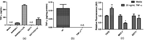
E0771 breast cancer cells do not produce nor respond to TNF-. (a): In vitro TNF- production: Supernatants from E0771 tumor cells showed no detectable TNF- in vitro (E0771 bar). Supernatants from TAMs isolated from E0771 tumors, in contrast, did produce detectable TNF- ( bar). To confirm sensitivity of the assay, RAW264.7-transformed murine macrophages produced detectable TNF- both when unstimulated (RAW264.7 bar) and upon activation with LPS for 24 h (RAW264.7+LPS bar). TNF- ELISA sensitivity was , samples for all but , where . Both the media alone control (media bar) and E0771 supernatants (E0771 bar) registered below sensitivity (not detectable). (b): In vivo TNF- production: also by this same ELISA assay, TNF- was detectable in E0771 tumors grown in C57Bl/6 mice (wt bar) but not in mice lacking TNF- [TNF- bar]. (c): Proliferation responses to TNF-: three breast tumor cell lines (T47D, MCF-7, and E0771) were treated with media control or TNF-. Pairwise comparisons indicate that T47D proliferation was significantly elevated by TNF- at 48 h (), MCF-7 proliferation was significantly decreased (), whereas only E0771 proliferation was unchanged in response to TNF-. Proliferation was assessed by fluorescent intensity of CyQuant DNA-binding dye standardized to that of cells in untreated media, per group.