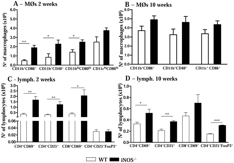
Figure 2. iNOS activity controls the influx of activated mononuclear phagocytes and T cells to the lungs.
Flow cytometry characterization of lung infiltrating leucocytes (LIL) from iNOS−/− and WT mice after i.t. infection with 1×10⁶ P. brasiliensis yeast cells. Lungs of iNOS−/− and WT mice (n = 6–8) were excised, washed in PBS, minced, and digested enzymatically. At weeks 2 (A, C) and 10 (B, D) after infection, lung cell suspensions were obtained and stained as described in Materials and Methods. (A, B) Phenotypic characterization of CD11b+ mononuclear phagocytes expressing CD80, CD86 and CD40 and dendritic cells expressing high levels of CD11c and CD86 (CD11chighCD86high). An increased presence of mononuclear phagocytes was observed in deficient mice, while the number of dendritic cells was equivalent in the lungs of both mouse strains. (C, D) Characterization of T cell subsets by flow cytometry in LIL obtained at weeks 2 (C) and 10 (D) after infection. To characterize the expansion of regulatory T cells in LIL, surface staining of CD25+ and intracellular FoxP3 expression were back-gated on the CD4+ T cell population. The acquisition and analysis gates were restricted to macrophages (A, B) or lymphocytes (C, D). The data represent the mean ± SEM of the results from 6–8 mice per group and are representative of two independent experiments. * (P<0.05), ** (P<0.01) and *** (P<0.001), compared with WT mice.

