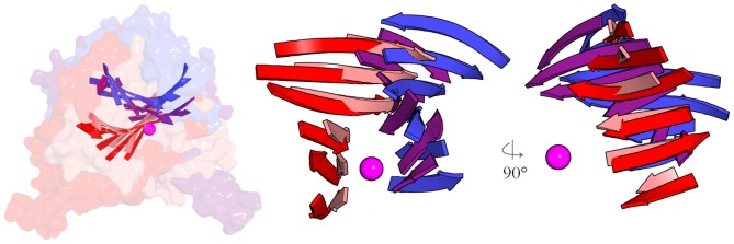
Figure 4. Retaining and inverting enzymes are entirely orthogonal.

Theβ-sheets of the metal-nucleotide-sugar binding GT-A foldsof glycosyltransferase structures are superimposed by centering on the metal ion (magenta sphere) and the coordinated phosphates reveal that the general architecture of entire inverting or retaining enzymes are skewed by ∼90°. Color coding: purple, inverting GlcAT-I; blue, inverting GalT1; red, retaining GTA; pink, retaining LgtC. Left panel shows the superimposed solvent-accessible surfaces of the four structures with the folds embedded; the right panels isolate the β-sheets and show orthogonal perspectives.
