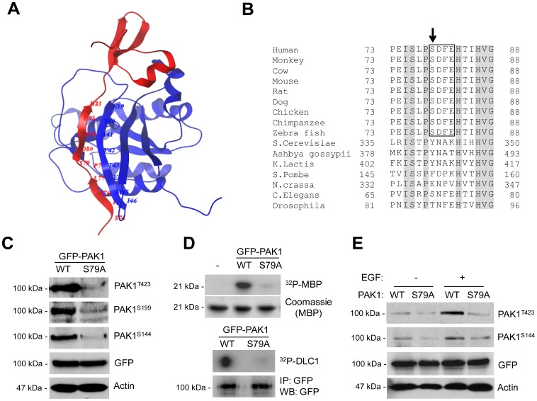
Figure 1. S79 within the CRIB domain is crucial for autophosphorylation and kinase activity of PAK1.
Overall Structures of CRIP domain of PAK1 and Sequence Comparisons. (A) Ribbon diagram showing a structural overview of CRIP domain of PAK1 (red) with bound Cdc42 (blue). Data Bank ID number 1E0A and visualized using Molsoft ICM Browser (Molsoft, L.L.C., San Diego, CA, USA). (B) Multiple sequence alignment of the amino acid sequence of PAK1 from 17 species. (C) Western blot analysis of phosphorylation of T423, S199, and S144 and of GFP-PAK1WT or GFP-PAK1S79A. (D) The kinase activity of PAK1 (GFP-PAK1WT or GFP-PAK1S79A) was assessed by in vitro phosphorylation assay using MBP (upper) or DLC1 peptide (lower) as substrate. (E) Western blot analysis of the effect of S79A mutation on the PAK1 autophosphorylation using anti-T423 and anti-S144 phospho-specific PAK1 antibodies. Cells were unstimulated (−) or stimulated (+) by EGF (100 ng/ml).

