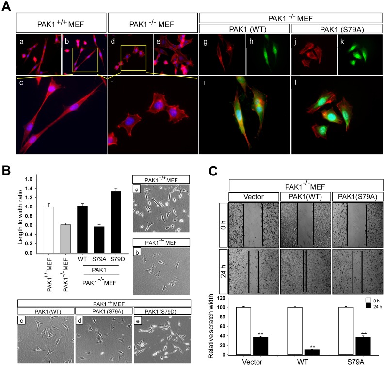
Figure 3. The S79A mutation impairs the ability of PAK1 to regulate cell morphology and motility.
(A) Wild type (a–c) and PAK1−/− (d–f) MEF cells were stained with the high affinity F-actin probe Phalloidin (red) and DAPI (blue). PAK1−/− MEF cells expressing GFP-PAK1WT (WT, g–i) and GFP-PAK1S79A (S79A, j–l) were stained with Phalloidin (red) and DAPI (blue) and visualized by GFP fluorescence (green). (B) Quantification of the length and width (L/W) ratio of MEF cells was obtained as described previously [44]. (C) Wound healing migration assays of PAK1−/− MEF cells infected with lentivirus expressing the vector control, GFP- PAK1WT, or GFP-PAK1S79A. Results were expressed as the percentage of the remaining area determined by normalizing the area of wound after 24 h to the initial wound area at 0 h (set to 100%). Each bar represents the mean ± S.D of five fields measured.

