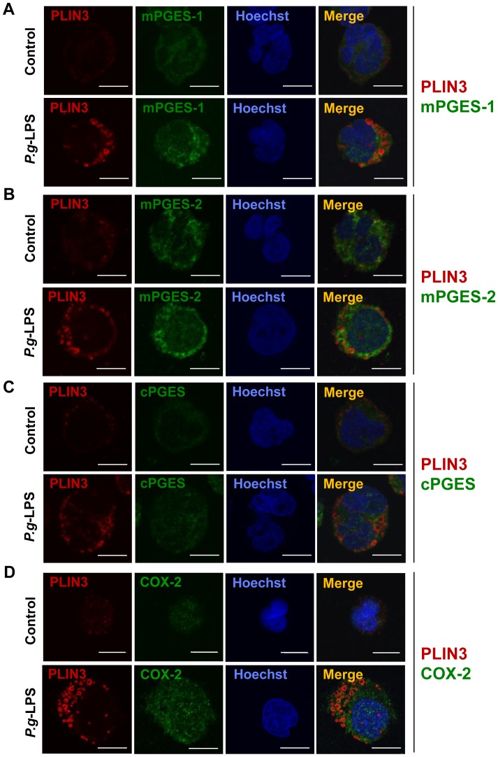
Figure 3. PLIN3 and PGE2 synthesizing enzymes induced in the P.g-LPS-treated HL-60 neutrophils distribute differently.
Differentiated HL-60 cells were treated with or without 10 µg/mL P.g-LPS for 12 h. Cells were fixed and then double stained with anti-PLIN3 and either anti-mPGES-1, anti-mPGES-2, anti-cPGES or COX-2 pAbs. Nuclei were stained with Hoechst33258. The cells were observed under confocal laser microscopy.

