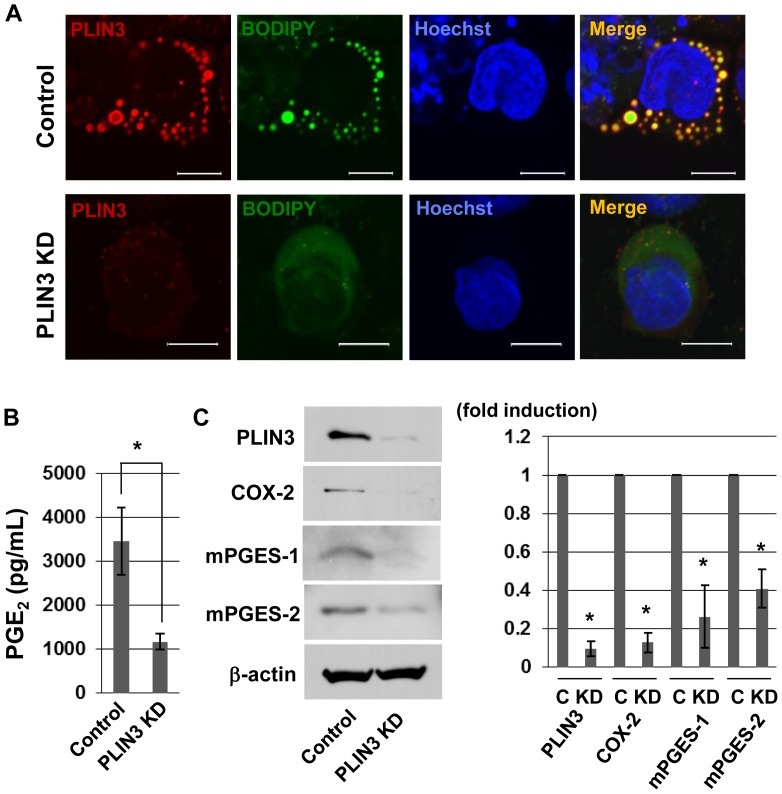
Figure 4. PLIN3 knockdown in HL-60-derived neutrophils suppressed the formation of LDs and production of PGE2.
A: After treatment of HL-60-derived neutrophils with PLIN3 siRNA or control siRNA for 72 h, the cells were stimulated with 10 µg/mL P.g-LPS for 12 h. The cells were fixed and labeled with BODIPY493/503 and anti-PLIN3 pAb. Bar = 10 mm. B: Cells were treated as described for A, and the PGE2 released into the media was measured by EIA. C: Whole cell lysates were subjected to SDS-PAGE and the protein levels of mPGES-1, mPGES-2, COX-2 and β-actin were analyzed by Western blotting. The band intensities were calculated by ImageJ software. Data are the mean ± SD of three independent experiments. *P<0.05.

