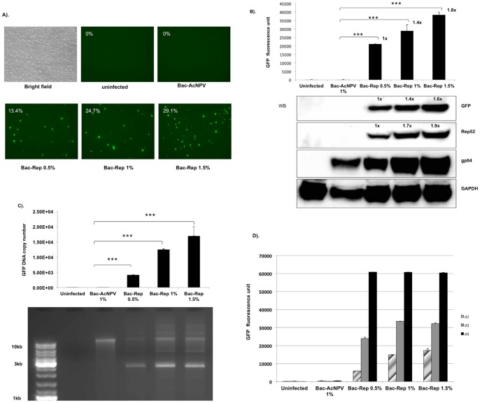
Figure 2. AAV Rep protein-dependent expression of GFP.
(A) Induction of GFP expression in blasticidin-S resistant Sf9/ITR-GFP cells in response to Bac-Rep infection. The clonal Sf9/ITR-GFP cell line contains a stably integrated copy of pFBGR-bsd. Uninfected cells or cells infected with wild-type baculovirus (Bac-AcNPV) lack detectable GFP expression as determined by FACS analysis. (upper row, 0% GFP positive cells). Addition of Bac-Rep inoculum (0.5%, 1.0%, and 1.5%; v:v) resulted in a dose-dependent increase in the number of GFP-positive cells (lower row). Images were obtained 3 days after infection. Magnification, 10x objective used for all images. (B) Quantitative analysis of GFP induction as a function of Bac-Rep dose. Cells exposed to increasing doses of Bac-Rep were harvested on day 3 post-infection. Fluorescent emission intensities were assessed from equivalent amounts of cell lysates (80 µg protein), using the fluorescent reader function of a real-time thermocycler (excitation 450–490 nm; emission 515–530 nm) (upper panel), *** indicates t-test significance (p<0.001). The relative fold-increase in GFP fluorescence is indicated by the values next to each bar. Protein concentrations were determined in the lysates and approximately 120 µg of each sample was fractionated electrophoretically using SDS-polyacrylamide gels. Proteins were electroblotted onto nitrocellulose membranes for western blot detection of GFP, Rep52, gp64, and GAPDH (used as a protein loading and transfer standard, lower panel). Relative levels of protein are indicated by the values above the protein band. (C) Analysis of the increase of GFP-vector DNA in response to Bac-Rep infection. Low molecular weight DNA was recovered from the cells and the quantity of GFP vector genomes determined using real-time qPCR (upper panel). Both the uninfected cell control and the wild-type baculovirus-infected cell control lysates produced a relatively low PCR signal (150 and 175 copies per cell, respectively). The GFP vector DNA copy number increased to 21087 copies per cell with 0.5% (v:v) Bac-Rep. At 1% (v:v) Bac-Rep, the copy number increased to 28862 copies per cell and 1.5% (v:v) Bac-Rep increased the copy number to 38286 per cell. The statistical significance was determined by Student's t-test. ***, p<0.001. Lower panel – The samples were analyzed by agarose-ethidium bromide gel electrophoresis (1% agarose). No detectable CELiD DNA was recovered from uninfected cells. A band >10 kb appears in lysates from all baculovirus-infected cells. (D) Time-course of GFP fluorescence. Sf9/ITR-GFP cells were inoculated with either wtBac (Bac-ACNPV) or Bac-Rep (0.5%, 1%, and 1.5% (v:v)). GFP fluorescence was measured for the indicated cellular lysate at 1, 2, and 3 days post-infection.

