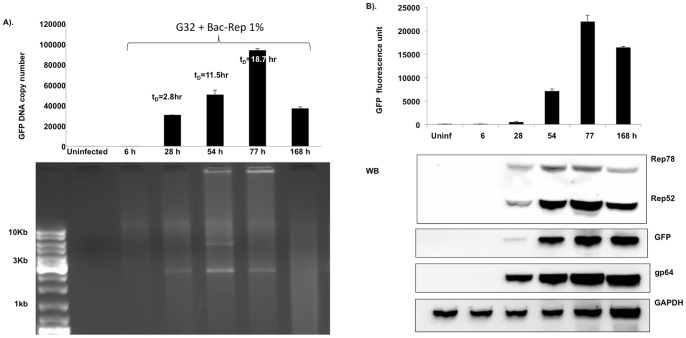
Figure 3. Rate of GFP vector DNA accumulation.
(A). Cells were inoculated with 1% (v:v) Bac-Rep stock and sampled at 6, 28, 54, 77, and 168 hr post-infection. The GFP-vector DNA content was determined by qPCR (upper panel). The doubling time of the GFP vector DNA was determined using the following algorithm: tD = (t2–t1) log 2/log (t2/t1), where tD is the doubling time and tn is the GFP copy number at a given time point. The tD (28 hr) = 2.8 hr, tD (54 hr) = 11.5 hr, and tD (77 hr) = 18.7 hr. (B) Time course of protein expression in Sf9/ITR-GFP cells. GFP fluorescence was measured in cell lysates obtained from the time-course described in (A) (upper panel). Aliquots from each time point were fractionated on SDS-polyacrylamide gels and transferred to nitrocellulose membranes for western blot analysis to detect Rep proteins, GFP proteins, gp64, and GAPDH (used as a loading and transfer control).

