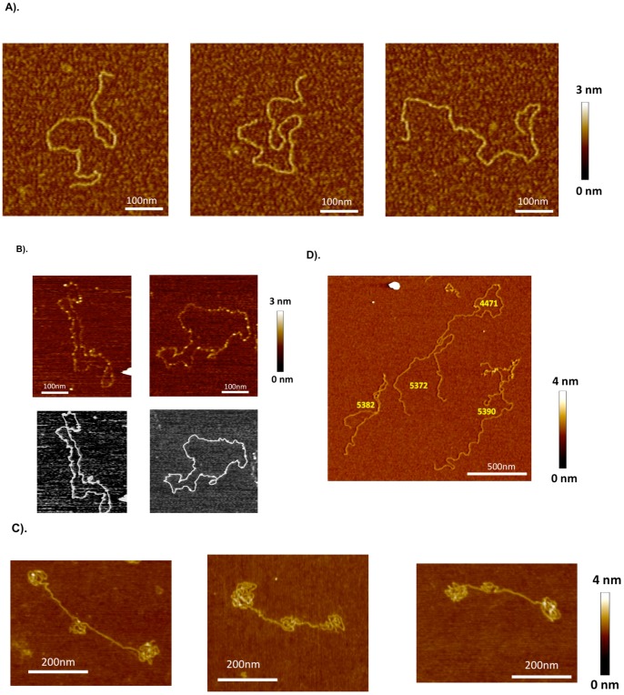
Figure 6. AFM images of CELiD vector DNA chains adsorbed onto APS-treated mica.
(A) Typical images of the CELiD-GFP monomer deposited from aqueous solution (dH2O). The monomer is indistinguishable from standard double-stranded, linear DNA. All images are 450×450 nm. (B) CELiD-GFP monomers adsorbed onto mica substrate immediately after being exposed to denaturing conditions. The height of the chain is about half that of the native monomer chain confirming that the loops are single-stranded. These monomers are closed loops with randomly located, condensed regions (brighter spots along the loops). The lengths of these loops are consistent with denatured monomers supporting the model of a covalently closed, duplex conformation under native conditions. Top images are of the denatured monomers. Bottom images show the corresponding traced loops. (C) Images of individual CELiD-GFP dimers adsorbed from H2O. The chains are double-stranded, linear DNA with lengths twice that of the monomer and exhibit a characteristic conformation with three condensed regions, one located centrally and the other two at opposite ends of the molecule. (D) Under high salt conditions (0.5 M NaCl), the condensed regions of the CELiD-GFP dimer relax and the chains take on conformations typical of double-stranded, linear DNA with length twice that of the monomer CELiD vector DNA. Numbers in yellow indicate the chain length in bp, estimated by path tracing.

