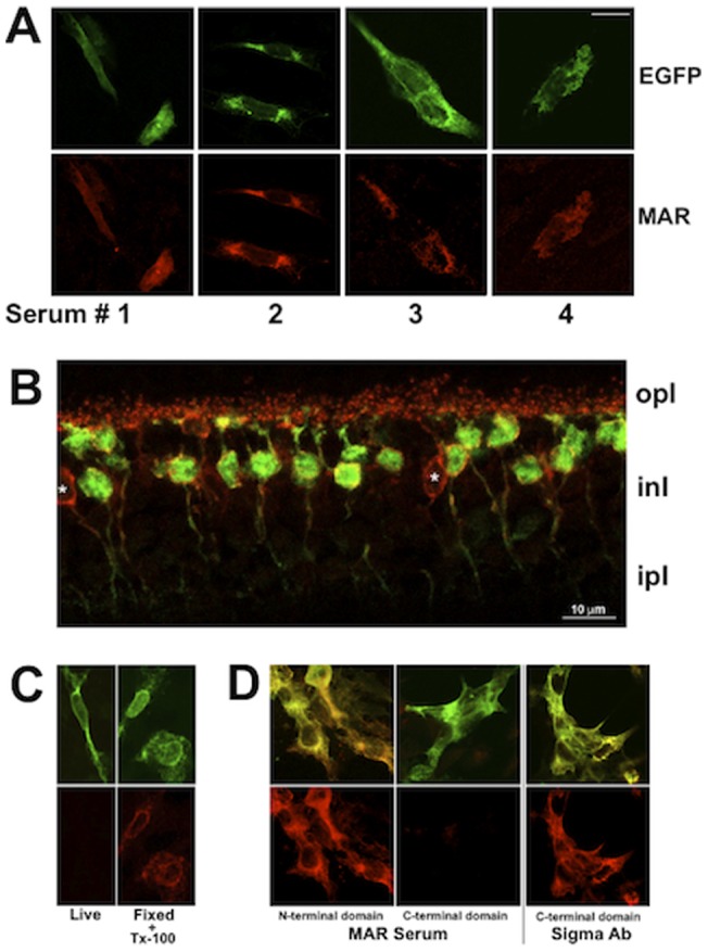
Figure 1. Identification of TRPM1-immunoreactive MAR sera.

A) MAR sera labeling of CHO cells transfected with EGFP-mTRPM1. EGFP is shown in green and the MAR serum immunoreactivity in red. B) Double immunofluorescence labeling of a mouse retina section with MAR serum #2 (red) and anti-PKC(green). Putative cone ON-bipolar cells are marked with asterisks. Scale bars = 10 µm. Abbreviations: OPL, outer plexiform layer; INL, inner nuclear layer; IPL inner plexiform layer. C) CHO cells transfected with EGFP-mTRPM1 were not immunolabeled when incubated with TRPM1-positive MAR serum while alive (left panels). TRPM1 immunofluorescence is revealed by applying the MAR serum after fixing and permeabilizing the cells (right panels). Top images: EGFP, bottom images: MAR immunofluorescence. D) Immunofluorescent labeling of N- and C-terminal TRPM1 peptides with MAR serum #2 (left) or a commercial anti-TRPM1 antibody against the C-terminus (Sigma Ab) as positive control (right). Top row: Superimposition of EGFP (green) and immunofluorescence (red) with co-localization appearing yellow. Bottom row: TRPM1 immunofluorescence.
