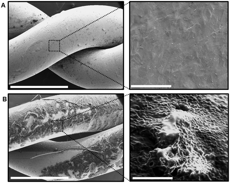
Figure 1. Scanning electron microscopy images of infected stainless steel wires used for sternotomy closure.
(A) Left panel is a SEM at 60x magnification of an unused sterile stainless steel wire, twisted in a way similar to that after sternotomy closure. Scale bar = 1 mm. Right panel is a higher magnification (10,000x) of the dashed box area in the left panel, showing the metal surface of the wire. Scale bar = 5 µm (B) Left panel is a SEM at 60x magnification of stainless steel wire after overnight incubation with MRSA strain USA300. Scale bar = 1 mm. Note the wire metal surface is coated by a film of material. Right panel is a higher magnification (10,000x) of the dashed box area in the left panel, showing clusters of cocci attached to the extracted wire and embedded within amorphous slime. Scale bar = 5 µm.

