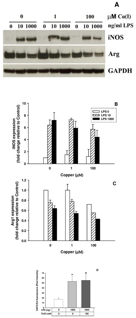
Figure 4. Copper does not inhibit expression and activity of iNOS and Arginase I in LPS-treated BV2 microglia.

BV2 cells were challenged with 1 or 100 μM Cu(I) and 8 hrs later stimulated with 0, 10 or 1000ng/mL LPS. Cell suspensions were collected 24 hrs after LPS challenge and an equal amount of protein measured for iNOS and Arginase I with GAPDH as internal control by western blot (A). Densitometry analysis of iNOS and Arginase was normalized by GAPDH as total protein loading (B and C). The effects of Cu(I) on intracellular NO content in LPS-treated BV2 microglia are shown in D. Cells were challenged with 100 μM Cu(I) and, 8 hrs later, stimulated with 1000 ng/mL LPS. Six hrs after LPS treatment, the cells were loaded with DAF-2DA for 30 min. * represents LPS significantly different from Control, p=0.003, and # represents Control significantly different from LPS 1000 ng/mL & Cu(I) 100 μM, p=0.0001.
