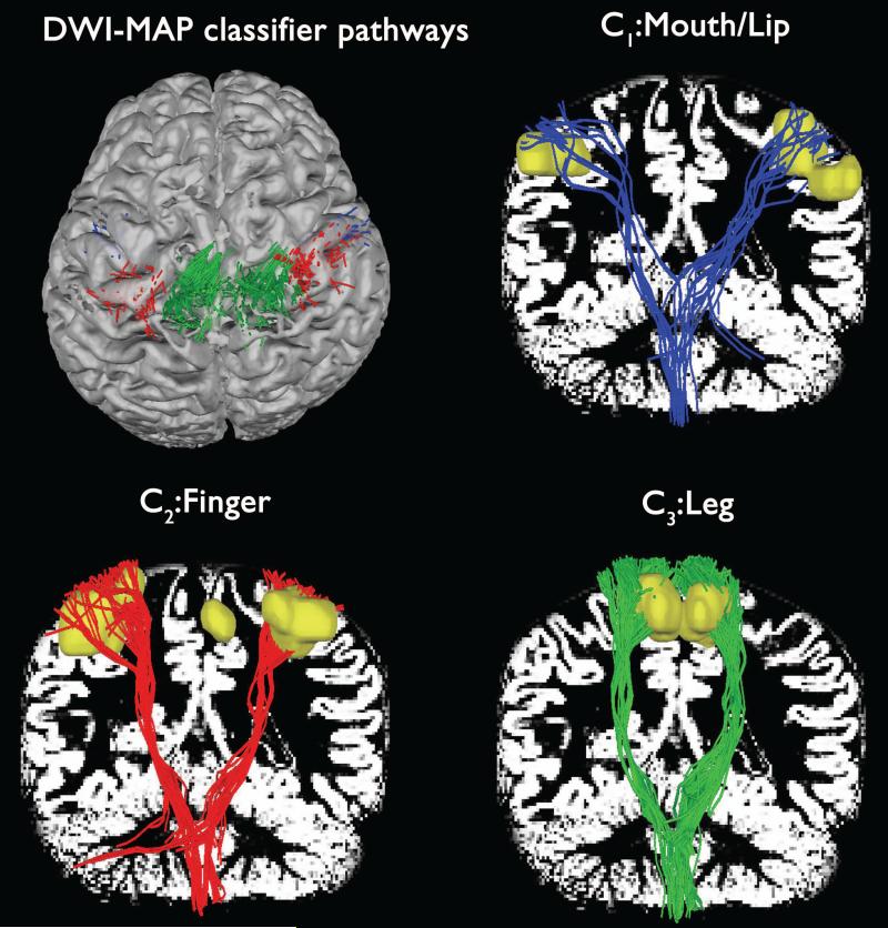
Figure 2.
Automatic detection of three pathways using the DWI-MAP classifier obtained from a normal participant. To demonstrate the reliability of the MAP classification, three pathways, C1: mouth/lip (blue), C2: finger (red), C3: leg (green) were presented with corresponding fMRI activations (yellow clusters). White background shows gray matter map.

