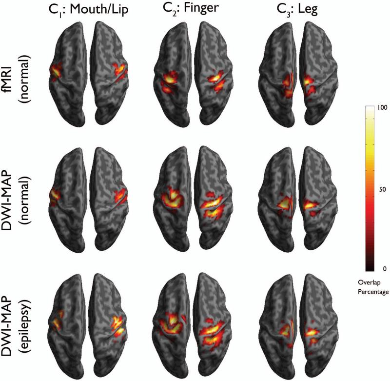
Figure 4.
Comparison of overlap percentage maps obtained from fMRI in healthy children (n=17, top), DWI-MAP classification in healthy children (n=17, middle), and DWI-MAP classification in children with epilepsy (n=20, bottom). Color bar indicates the overlap percentage of all cortical localizations obtained from each group.

