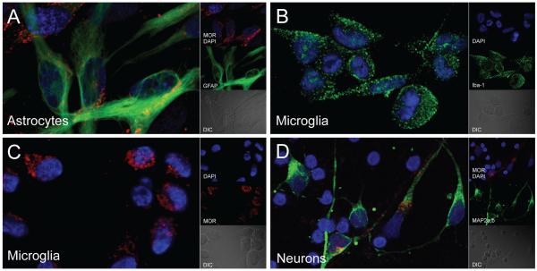
Fig. 1.
Appearance of MOR across human CNS cell types. Representative images of primary human a astrocytes, b, c microglia, and d neurons immunolabeled for the N-terminus of MOR (red) and their respective cell-type specific markers (green) (GFAP, Iba-1, and MAP2a,b, respectively). DAPI (blue) staining indicates cell nuclei. DIC, differential interference contrast microscopy

