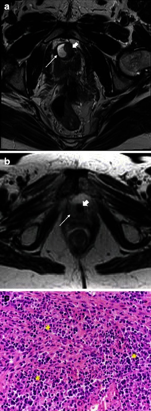
Fig. 11.

a Urethral tumour mimics: Complex/inflamed diverticulum mimicking a mass. Axial T2-weighted image shows a high signal collection (arrow) along the right margin of the urethra (block arrow), in keeping with an uncomplicated urethral diverticulum. b Urethral tumour mimics: complex/inflamed diverticulum mimicking a mass. In contrast, axial T2-weighted image in a different patient demonstrates an inflamed diverticulum as intermediate signal (arrow) along the right margin of the urethra (block arrow). c Urethral tumour mimics: complex/inflamed diverticulum mimicking a mass. Histological section shows an intense acute and chronic inflammatory infiltrate throughout the image (*), with a mix of neutrophils, lymphocytes and monocytes. (Haematoxylin and eosin, original magnification 400×)
