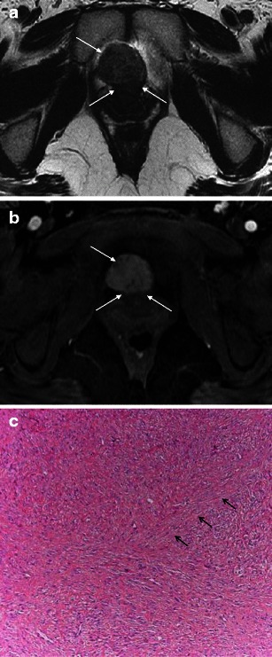
Fig. 7.

a Benign lesions: urethral leiomyoma. Axial T2-weighted image shows an exophytic, well demarcated mass arising from the urethra anteriorly (arrows). b Benign lesions: urethral leiomyoma. Axial T1-weighted image after intravenous gadolinium-based contrast administration demonstrates homogeneous enhancement of the mass (arrows). c Benign lesions: urethral leiomyoma. Histological specimen shows characteristic intersecting fascicles of smooth muscle throughout the image in this benign stromal tumour. The arrows point to one of these fascicle bands. (Haematoxylin and eosin, original magnification 100×)
