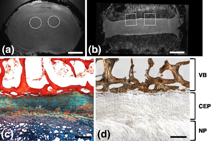
Fig. 4.
MRI and histology images of the same specimen (63 years male, L2L3, Grade 2.6). Axial (a) and coronal (b) FLASH MRI of the whole disc, showing approximate locations of biopsy punches used for histological analysis. c Representative histology section of the CEP stained with Alcian blue (glycosaminoglycans) and picrosirius red (collagen), showing adjacent NP and vertebral bone. d Von Kossa staining of an undecalcified section, showing regions of bone distinct from CEP and minimal CEP calcification. (Scale bars in a and b = 1 cm and in c and d = 0.5 mm; VB vertebral bone)

