Figure 3.
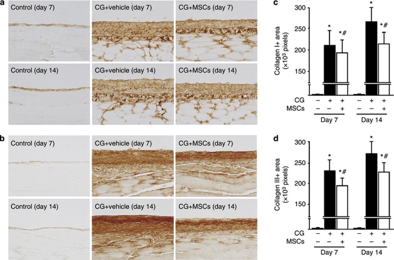
Mesenchymal stem cells (MSCs) suppressed collagen I and III expressions in rats with peritoneal fibrosis. Immunohistochemical analyses of (a) collagen I and (b) collagen III expression in peritoneal tissues on days 7 and 14 (original magnification × 200) in control rats, chlorhexidine gluconate (CG)-injected rats treated with the vehicle alone, and CG-injected rats treated with MSCs. (c, d) The numbers of collagen I+ and III+ pixels were increased on days 7 and 14 in CG-injected rats treated with the vehicle, whereas the numbers of collagen I+ and III+ pixels were significantly smaller on days 7 and 14 in CG-injected rats treated with MSCs. *P<0.01 versus control group, #P<0.05 versus CG+vehicle group.

