Figure 9.
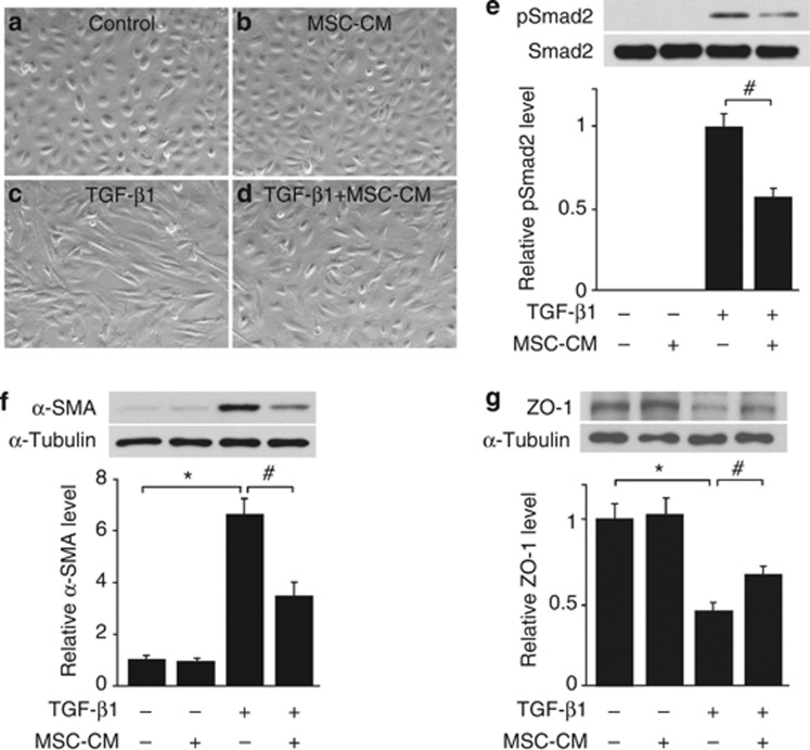
Mesenchymal stem cell (MSC)-conditioned media (CM) blocked transforming growth factor-β1 (TGF-β1)-induced epithelial-to-mesenchymal transition (EMT). Representative photomicrograph shows characteristic cobblestone-like appearances of (a) control human peritoneal mesothelial cells (HPMCs) and (b) HPMCs incubated with MSC-CM without TGF-β1 stimulation. Fibroblast-like change was observed following TGF-β1 stimulation for 48 h, but the change was reduced in (d) HPMCs incubated with MSC-CM than in (c) HPMCs incubated with normal medium. HPMCs were observed by an independent investigator who was blinded to the experimental conditions. (e) Western blot analysis shows that MSC-CM inhibited phosphorylation of Smad2 in HPMCs after 30 min of TGF-β1 stimulation. (f, g) TGF-β1 treatment for 48 h caused increased α-smooth muscle actin (α-SMA) protein expression and decreased zonula occludens-1 (ZO-1) protein expression. MSC-CM attenuated these TGF-β1-induced EMT responses (n=6). *P<0.01, #P<0.05.

