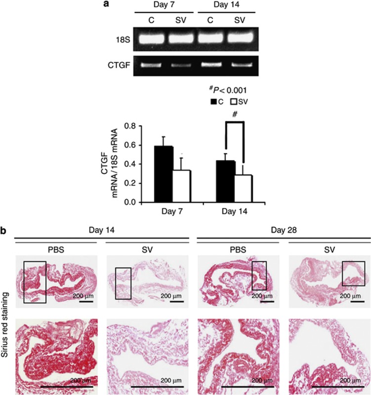
Figure 7.
Gene expression of connective tissue growth factor (CTGF) and Picrosirius red staining are reduced in simvastatin (SV)-treated vessels when compared with controls (Cs). (a) The real-time polymerase chain reaction (RT-PCR) analysis of CTGF expression after treatment with either C or SV at day 7 or day 14 after arteriovenous fistula (AVF) placement. A typical blot is shown in the upper panel and the pooled data in the lower panel. (a) The average CTGF expression is significantly decreased at day 14 in the SV-treated vessels when compared with Cs (P<0.001). (b) Representative sections after Picrosirius red staining at the venous stenosis after treatment with either C or SV at day 14 or day 28 after AVF placement. Upper panel: original magnification, × 40, and the lower panel is a magnification view of the box. The more intense red staining is representative of collagen 1 and 3 staining. Qualitatively, there is decreased Sirius red staining in the simvastatin-treated vessels when compared with Cs. Each bar represents mean±s.e.m. of 3–5 animals. Two-way analysis of variance (ANOVA) followed by Student's t-test with post hoc Bonferroni's correction was performed. Significant differences between the SV-treated group and controls are indicated by *P<0.01.

