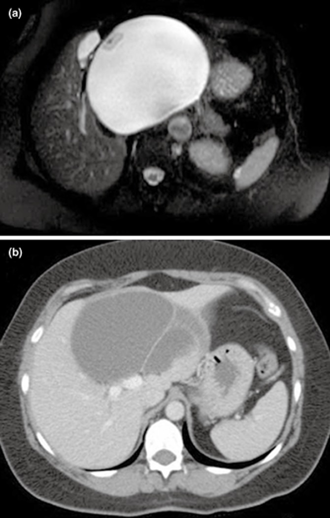
Figure 1.

(a) T2-weighted magnetic resonance imaging (MRI) demonstrating a 14 × 15 cm centrally-located liver cystadenoma with mural nodularity and inferior vena cava compression in a 63-year-old female. (b) Computed tomography (CT) scan demonstrating an 11 × 8 cm centrally-located multi-sepatated liver cystadenoma arising above the portal bifurcation in a 56-year-old female
