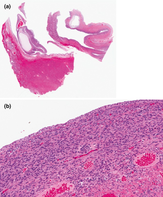
Figure 2.

(a) Mucinous cystadenoma (10×, haematoxylin and eosin stain). There is a large cyst lined by a simple biliary-type columnar epithelium. (b) The ovarian-like mesenchymal stroma is seen underneath the simple epithelium (100×, haematoxylin and eosin stain).
