
Figure 3. Alterations in nuclear expression of p53 and PGC-1α with acute exercise.
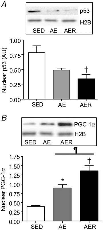
Nuclear content of p53 (A) steadily decreased, and that of PGC-1α (B) progressively increased in the AE and AER animal groups. Histone 2B was used as a loading control and did not change during the conditions. *P < 0.05 AE vs. SED, †P < 0.05 AER vs. SED. ¶P < 0.05 AER vs. AE. Data are presented as mean ± SEM.