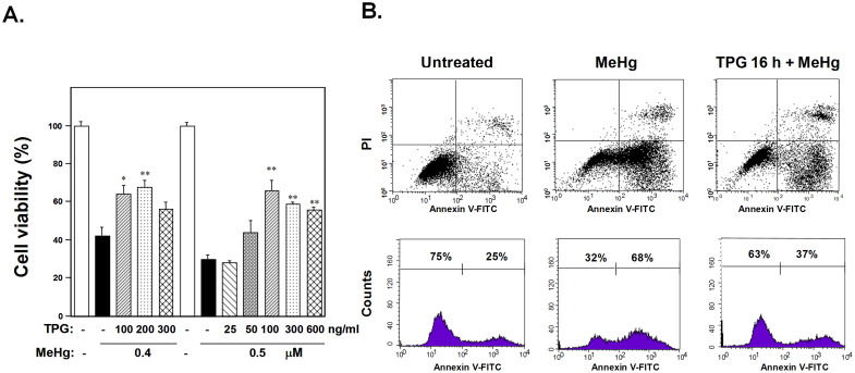
Figure 1. Effect of pretreatment with TPG on MeHg cytotoxicity.
(A) Cell viability of C2C12-DMPK 160 cells pretreated with TPG 16 h before exposure to 0.4 or 0.5 μM MeHg was determined. Pretreatment with TPG (100–300 ng/ml) attenuated MeHg cytotoxicity. The viability of untreated cells was regarded as 100%. Values represent means ± SE (n = 6). *, **Significantly different from TPG-untreated and MeHg-treated cells by a one-way ANOVA (*p < 0.05, **p < 0.01). (B) Apoptosis analysis. The upper panel shows flow cytometry analysis of C2C12-DMPK160 stained with propidium iodide (PI) and FITC-Annexin V. The vertical axis indicates PI fluorescence intensity and horizontal axis Annexin V fluorescence. Exposure to 0.4 μM MeHg for 16 h increased the number of cells undergoing apoptosis (Annexin V-FITC-positive and PI-negative). A minor population of cells was observed to be Annexin V-FITC- and PI-positive, indicating that they were in end-stage apoptosis or already dead. The lower panel shows the profile of frequency of viable cells (Annexin V-FITC- and PI-negative) and cells undergoing apoptosis (Annexin V-FITC-positive and PI-negative). Pretreatment with TPG decreased the frequency of cells undergoing apoptosis.

