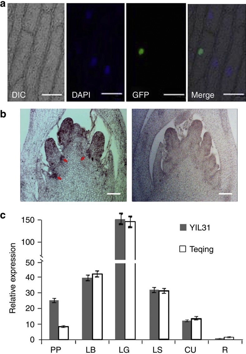
Figure 5. Subcellular localization and expression pattern of the OsLG1 gene.
(a) Subcellular localization of OsLG1. The OsLG1–GFP fusion gene under the control of the CaMV35S promoter was expressed transiently in onion epidermal cells. Scale bars, 100 μm. (b) OsLG1 gene expression revealed by mRNA in situ hybridization in the young panicle of YIL31. Arrowheads indicate OsLG1 expression in the panicle pulvinus. Left, antisense probe; right, sense probe (control). Scale bars, 100 μm. (c) Expression of OsLG1 in different tissues of YIL31 and Teqing by quantitative RT–PCR at the late panicle development stage. PP, panicle pulvinus; LB, leaf blade; LG, leaf ligule; LS, leaf sheath; CU, culm; R, root. Values are means and s.d. of three independent experiments.

